Skeletal Muscle Physiology
Skeletal muscle generates force to produce movement.
It works with bones and joints to allow voluntary control of body position and motion.
Key point: Skeletal muscle = voluntary + striated + force generation
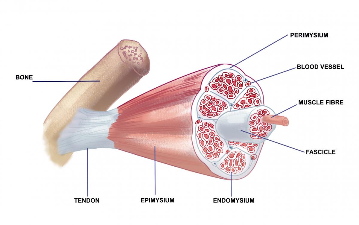
Skeletal muscle is organised in layers from whole muscle to cellular level.
- Whole muscle → epimysium
- Fascicles (bundles of fibres) → perimysium
- Individual muscle fibres → endomysium
Muscle fibres are long, multinucleated cells arranged in parallel.
Exam rule:
- Whole muscle → epimysium
- Fascicle → perimysium
- Muscle fibre → endomysium
Muscle Fibre Structure
Each muscle fibre is surrounded by the sarcolemma.
The sarcolemma is the cell membrane and forms invaginations called T-tubules.
T-tubules conduct electrical signals from the surface into the interior of the muscle fibre.
Inside each fibre are myofibrils, which run along the length of the cell.
Sarcomere: The Functional Unit
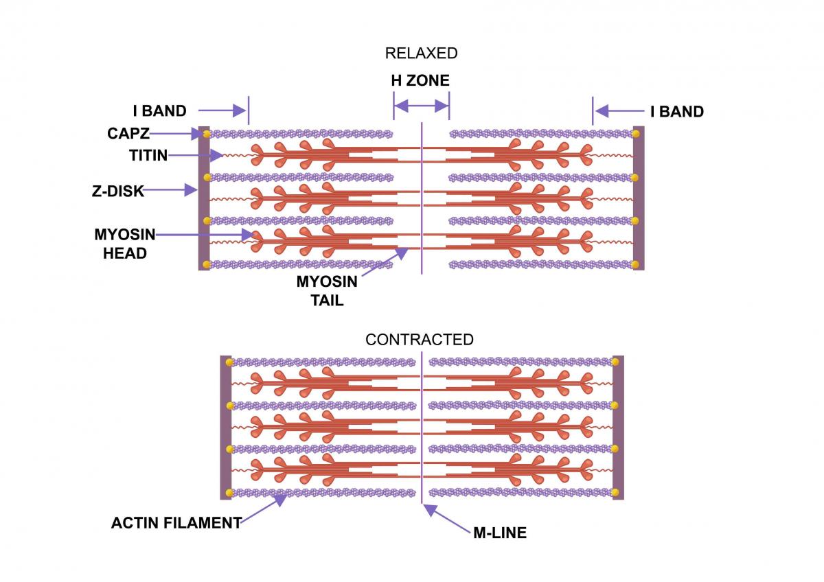
The sarcomere is the basic unit of contraction.
It lies between two Z-lines.
It contains organised filaments:
- Thin filaments → actin
- Thick filaments → myosin
- Elastic filaments → titin
Key regions:
- A-band → length of myosin
- I-band → actin only
- H-zone → myosin only
- M-line → central support
Exam rule:
- Thin filament → actin
- Thick filament → myosin
Mechanism of Contraction
Muscle contraction occurs via the sliding filament mechanism.
Myosin heads bind to actin to form cross-bridges.
This pulls actin filaments towards the centre of the sarcomere.
This shortens the sarcomere and generates force.
Changes During Contraction (High Yield)
During contraction, filaments do not shorten.
Actin slides over myosin, increasing overlap.
This produces predictable structural changes:
- A band → remains constant
- I band → decreases in length
- H zone → decreases and may disappear
- Z lines → move closer together
Key point: Myofilaments do not shorten; they slide past each other
Exam rule:
- A band = constant
- I band + H zone = decrease
Excitation–Contraction Coupling
Contraction begins with a nerve impulse.
At the neuromuscular junction:
- Acetylcholine is released
- Binds to ligand-gated sodium channels
- Causes depolarisation of the muscle membrane
The action potential travels along the sarcolemma and into the cell via T-tubules. This ensures rapid and uniform depolarisation throughout the cell.
This triggers calcium release from the sarcoplasmic reticulum. Calcium binds to troponin.
This exposes actin binding sites and allows cross-bridge formation.
Exam rule:
- Ca²⁺ binds → troponin
- T-tubules → carry action potential into muscle fibre
Role of ATP
ATP is required for contraction:
- Allows myosin to detach from actin
- Provides energy for the power stroke
Without ATP, contraction cannot cycle.
Length–Tension Relationship (High Yield)
The force generated by a muscle depends on the initial sarcomere length.
Maximal tension occurs when there is optimal overlap between actin and myosin.
If the sarcomere is too stretched:
- Reduced overlap
- Fewer cross-bridges
- Reduced force
If the sarcomere is too shortened:
- Excessive overlap
- Interference with cross-bridge formation
- Reduced force
Key point: Maximal force = optimal actin–myosin overlap
Motor Units and Force Generation
A motor unit consists of one motor neuron and its muscle fibres.
Force is increased by recruiting more motor units.
Greater recruitment → greater force.
Key point: Force depends on motor unit recruitment
Clinical Relevance
These principles explain common clinical findings.
- NMJ dysfunction → weakness with normal muscle structure
- Reduced calcium release → reduced contraction
- ATP depletion → impaired relaxation
Typical presentation:
- Muscle weakness → reduced force generation
Key Exam Tips
Skeletal muscle questions focus on structure and sequence.
In every question, focus on:
- Filament type
- Calcium role
- Level of organisation
Common traps to avoid:
- Confusing connective tissue layers
- Mixing up actin and myosin roles
- Forgetting that troponin binds calcium
- Confusing optimal with maximum overlap
Exam rule: Muscle contraction = Ca²⁺ + troponin + actin–myosin interaction