The heart produces a symphony of sounds that can provide valuable insights into a patient’s cardiovascular health. This article will delve into the fundamental principles of heart sounds, their physiological basis, and their clinical significance, equipping medical professionals with the knowledge to decode this vital symphony.
An audio example of the normal heart sounds can be heard here (courtesy of Littman stethoscopes).
The physiology of the heart sounds
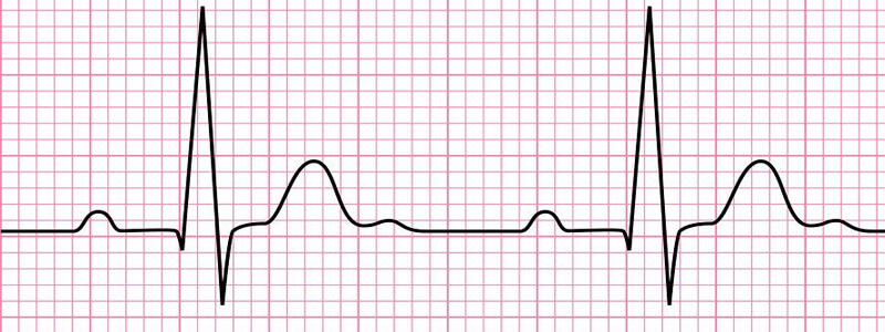
The first step in unravelling the symphony of the heart lies in comprehending its physiological basis. The cardiac cycle, composed of systole and diastole, generates two distinct heart sounds: S1 and S2. S1, the first heart sound, is caused by the closure of the atrioventricular (AV) valves, primarily the mitral and tricuspid valves, and marks the beginning of systole. S2, the second heart sound, arises from the closure of the semilunar valves, particularly the aortic and pulmonic valves, signifying the onset of diastole.
The first heart sound
The first heart sound (S1), sometimes described as the “lub” sound, is a crucial component of the heart’s symphony. It is generated by the abrupt cessation of blood flow between the atria and ventricles during systole. It is helpful to remember that S1 coincides with the carotid pulse to help differentiate it from other heart sounds.
The closure of the AV valves produces a low-pitched sound that can be best heard at the apex of the heart. It corresponds with the end of diastole and the beginning of ventricular systole and precedes the upstroke of the carotid pulsation.
The following conditions are associated with a loud S1:
- Increased transvalvular gradient (e.g. mitral stenosis, tricupsid stenosis)
- Increased force of ventricular contraction (e.g. tachycardia, hyperdynamic states such as fever and thyrotoxicosis)
- Shortened PR interval (e.g. Wolff-Parkinson-White syndrome)
- Mitral valve prolapse
- Thin individuals
The following conditions are associated with a soft S1:
- Inappropriate apposition of the AV valves (e.g. mitral regurgitation, tricuspid regurgitation)
- Prolonged PR interval (e.g. heart block, digoxin toxicity)
- Decreased force of ventricular contraction (e.g. myocarditis, myocardial infarction)
- Increased distance from the heart (e.g. obesity, emphysema, pericardial effusion)
The second heart sound
Following S1, the cardiac symphony progresses to the second heart sound, S2, which manifests as the “dub” sound. S2 occurs due to the closure of the aortic and pulmonic valves at the onset of diastole. This sound is relatively higher in pitch and is best appreciated at the base of the heart. It is important to note that S2 is divided into two components: A2, corresponding to the closure of the aortic valve, and P2, denoting the closure of the pulmonic valve. Splitting during inspiration is a normal finding.
The following conditions are associated with a loud S2:
- Systemic hypertension (loud A2)
- Pulmonary hypertension (loud P2)
- Hyperdynamic states (e.g. tachycardia, fever, thyrotoxicosis)
- Atrial septal defect (loud A2)
The following conditions are associated with a soft S2:
- Decreased aortic diastolic pressure (e.g. aortic regurgitation)
- Poorly mobile cusps (e.g. calcification of the aortic valve)
- Aortic root dilatation
- Pulmonary stenosis (soft P2)
The following conditions are associated with a widely split S2:
- Deep inspiration
- Right bundle branch block
- Prolonged right ventricular systole (e.g. pulmonary stenosis, P.E.)
- Severe mitral regurgitation
- Atrial septal defect (fixed splitting, doesn’t vary with respiration)
The following conditions are associated with reversed splitting of S2 (paradoxical splitting with P2 occurring before A2):
- Deep expiration
- Left bundle branch block
- Prolonged left ventricular systole (e.g. severe aortic stenosis, hypertrophic cardiomyopathy)
- Severe aortic stenosis
- Right ventricular pacing
- Wolff-Parkinson-White (type B)
Extra heart sounds
Beyond the fundamental S1 and S2 sounds, you should also be aware of additional heart sounds that may indicate underlying pathology. These include S3 and S4, referred to as the third and fourth heart sounds, respectively.
The third heart sound
The third heart sound is a low-frequency, brief vibration that occurs in early diastole at the end of the rapid diastolic filling period of the right or left ventricle. It is associated with heart failure and volume overload but may also be a normal finding in children and young adults.
The genesis of the 3rd heart sound is controversial. Possible explanations for the third heart sound include the impact of flowing blood against an incompliant ventricle, the impact of the ventricle against the inner chest wall or a sound originating within the ventricular apex due to sudden limitation of longitudinal expansion.
The fourth heart sound
The fourth heart sound is a rare extra sound that can be heard immediately before the two normal ‘lub-dub’ heart sounds. It occurs during late diastolic filling of the ventricle during atrial systole. It results from vibrations generated within the ventricles, and its presence usually indicates increased resistance to the filling of the left or right ventricle because of a stiff ventricular wall.
S4 can be associated with several conditions, including the following:
- Ventricular hypertrophy
- Aortic stenosis
- Post-MI ventricular fibrosis
- Hypertrophic cardiomyopathy
- Restrictive cardiomyopathy
Header image used on licence from Shutterstock