Trauma remains a leading cause of mortality worldwide, with chest injuries representing a significant portion of these cases. Whether resulting from motor vehicle accidents, falls, or penetrating trauma, injuries to the chest can lead to immediate compromise of vital functions, necessitating swift and accurate assessment and intervention.
The ATLS guidelines divide the chest injuries seen into trauma into two groups:
- Life-threatening injuries that should be identified and treated in the primary survey and;
- Potentially life-threatening injuries that should be identified and treated in the secondary survey
Overview of the life-threatening chest injuries (ATOM-FC)
The 6 ‘killer conditions’ that should be identified and treated in the primary survey can be remembered using the mnemonic ATOM-FC. These “killer conditions” can rapidly deteriorate a patient’s condition if left untreated, underscoring the importance of early recognition and prompt management in the pre-hospital and emergency department settings:
- Airway obstruction
- Tension pneumothorax
- Open pneumothorax
- Massive haemothorax
- Flail chest
- Cardiac tamponade
In addition, the following potentially life-threatening injuries should be identified and treated in the secondary survey:
- Simple pneumothorax
- Haemothorax
- Pulmonary contusion
- Tracheobronchial tree injury
- Blunt cardiac injury
- Traumatic aortic dissection
- Traumatic diaphragmatic injury
- Blunt oesophageal rupture
Airway Obstruction
An obstructed airway poses an imminent threat to ventilation and oxygenation, leading to hypoxia and respiratory failure.
Prompt assessment and clearance of the airway, along with interventions such as positioning, suctioning, or advanced airway management, are vital in ensuring adequate oxygen delivery to the patient’s lungs.
Tension Pneumothorax
A tension pneumothorax occurs when a ‘one-way valve’ air leak occurs from the lung or through the chest wall. There is a progressive build-up of air within the pleural space without any means of escape. This results in a progressive rise of pressure in the pleural space, which pushes the mediastinum into the opposite hemithorax. This can impede venous return to the heart and cause cardiovascular instability and cardiac arrest if untreated.
The following clinical features are characteristic of tension pneumothorax:
- Respiratory distress and cardiovascular instability
- Tracheal deviation away from the side of injury
- Unilateral absence of breath sounds on the side of injury
- Hyper-resonant percussion note on the side of injury
- Distended neck veins
- Cyanosis (late sign)
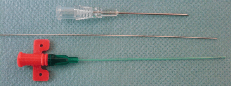
Tension pneumothorax is a clinical diagnosis and treatment should not be delayed for radiological confirmation. Treatment is with IMMEDIATE decompression via needle thoracocentesis.
Open Pneumothorax
Also known as a “sucking chest wound,” open pneumothorax occurs when there is a communication between the pleural space and the external environment. Once a chest wound is approximately two-thirds the diameter of the trachea or greater, air passes preferentially through the chest wall with each breath. This impairs effective ventilation, rapidly resulting in hypoxia and hypercarbia.
The following clinical features are characteristic of open pneumothorax:
- The presence of an open wound on the chest wall
- Respiratory distress and cardiovascular instability
- Reduced chest movement on the side of the injury
- Decreased breath sounds on the side of the injury
- Hyper-resonant percussion note on the side of the injury
- Bedside ultrasound scanning can be used to confirm the diagnosis rapidly
Emergency management involves sealing the wound with a three-sided dressing to form a ‘flutter-valve’. This allows air to escape the pleural cavity when the patient breathes out. The dressing will occlude when the patient breathes in, preventing air from entering. The wound will require formal exploration before closure, and a chest tube in a separate intercostal space should be sited as soon as possible.
Massive Haemothorax
A massive haemothorax results from the rapid accumulation of more than 1500 mL of blood or 1/3 of the patient’s blood volume in the chest cavity.
The classic signs of a massive haemothorax are:
- Decreased chest expansion
- Decreased breath sounds
- Dullness to percussion
The first step in the management of massive haemothorax should be the simultaneous restoration of blood volume and decompression of the chest cavity with a chest drain. The majority of haemothoraces will have stopped bleeding by the time management is commenced, and simple drainage is all that is required. All chest drains placed in trauma should be of sufficient calibre to drain the haemothorax without clotting (preferably 36F).
Flail Chest
Flail chest occurs when two or more adjacent ribs are fractured in two or more places, leading to a segment of the chest wall becoming detached from the rest of the thoracic cage. This results in a segment of the chest wall that is no longer in continuity with the rest of the thoracic cage. The ‘flail’ segment moves paradoxically to the rest of the chest, moving inwards on inspiration as the remainder of the chest expands and outwards on expiration as the remainder of the chest deflates.
The following clinical features are characteristic of flail chest:
- Respiratory distress and cardiovascular instability
- Paradoxical chest wall movement of the affected ‘flail’ segment
- Decreased breath sounds
- Bony crepitus on chest palpation
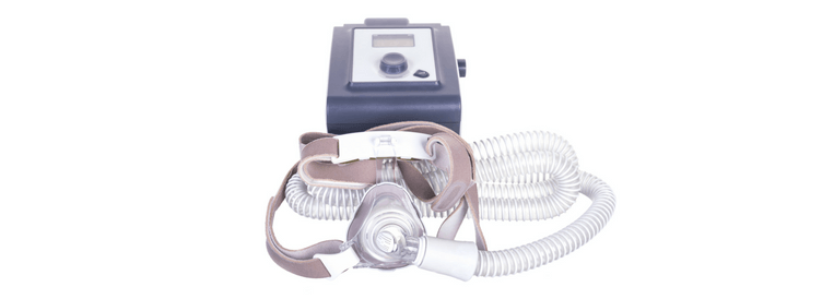
Initial management involves supplemental oxygen, analgesia, and chest splinting via direct pressure over the affected area. A chest tube may be necessary, and mechanical ventilation with PEEP is necessary in some cases that fail to improve with conservative treatment. Surgery can be performed to stabilise the displaced ribs if further intervention is required.
Cardiac Tamponade
Cardiac tamponade can occur following both penetrating and blunt thoracic trauma. The trauma causes the pericardial cavity to fill with blood from the heart, great vessels, or pericardial vessels.
The pericardial cavity (also referred to as the pericardial space) contains the lubricating pericardial fluid and is found between the parietal and visceral pericardium. The pericardial cavity usually contains around 20 ml of pericardial fluid. Once around 200 ml of fluid has accumulated, systolic dysfunction and impairment of cardiac output begin to develop. This is referred to as cardiac tamponade.
Cardiac tamponade can be recognised clinically by Beck’s triad of:
- Distended neck veins
- Muffled heart sounds
- Hypotension
The treatment of cardiac tamponade is urgent pericardiocentesis. Pericardiocentesis is the process by which excess fluid can be drained from the pericardial cavity. The procedure can be performed blind or under guidance by CT or ultrasound scan. The current consensus opinion is that it should only be carried out blindly in life-threatening circumstances.
Header image used on licence from Shutterstock